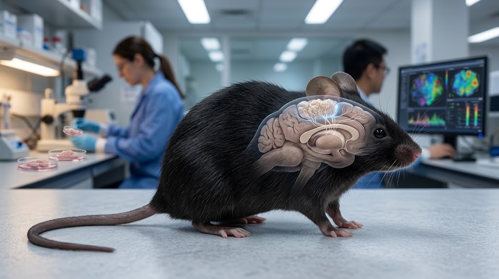
Stanford researchers created mice missing much of their cerebral cortex, then transplanted laboratory-grown human cortical organoids into the empty space. Three months later, human tissue made up more than 90% of the animals’ remaining cortical tissue by volume and had connected with the mouse nervous system. The experiment could transform brain-disease research—but it also raises an obvious question: how human can an animal brain become?
Published: September 17, 2026
Scientists Grew Human Brain Tissue Across Most of a Mouse Cortex—What Exactly Did They Create?
Human neurons grew, fired together, sent projections through the mouse nervous system and responded to injury. Yet the animals did not acquire a human brain, human intelligence or anything resembling human consciousness. The real breakthrough is subtler—and perhaps more important.
By History & Science Desk | September 17, 2026
A mouse was born missing much of the part of its brain associated with higher cognition.
Scientists then placed pieces of laboratory-grown human cortical tissue into the empty space.
The tissue survived.
It grew.
Human neurons connected with one another.
They extended nerve fibres into deeper parts of the mouse nervous system.
They displayed organized electrical activity.
And after three months, the transplanted human tissue made up more than 90% of the combined cortical tissue volume measured in the experimental animals.
That sentence sounds like the beginning of a science-fiction story.
It isn't.
It is a peer-reviewed experiment published this week in Nature by researchers led by Stanford neuroscientist Sergiu Pașca.
The animals are being called:
xenocortical mice.
And they may provide one of the most powerful new ways yet to study human brain development, cerebral palsy, epilepsy, autism, schizophrenia and other disorders that are extraordinarily difficult to recreate in ordinary laboratory animals.
But before imagining mice with human minds, an important correction is necessary.
The scientists did not transplant an intact human brain.
They did not create a miniature adult human cortex.
They did not demonstrate human consciousness inside a mouse.
And “90% human cortex” does not mean 90% of the mouse's entire brain became human.
What they created is both stranger and scientifically more useful.
Why Would Anyone Put Human Brain Tissue Into a Mouse?
Because the human brain is remarkably difficult to study while it is developing.
Researchers can examine donated adult brain tissue after death.
They can use brain scans.
They can study genes.
They can observe cells grown in dishes.
But scientists cannot experimentally manipulate a developing human cerebral cortex simply to see what happens.
That creates a huge problem.
Many neurological and psychiatric conditions begin during brain development long before symptoms become obvious.
Researchers therefore need models.
Mice have taught scientists an enormous amount about neuroscience.
But a mouse brain is not simply a small human brain.
Human cortical neurons mature much more slowly.
Some human brain-cell types differ from rodent cells.
Human developmental timing is different.
And certain disorders appear to involve biological features that ordinary mouse models reproduce poorly.
Scientists therefore began creating brain organoids.
What Is a Brain Organoid?
Take an adult human skin or blood cell.
Reprogram it into an induced pluripotent stem cell.
Then provide carefully controlled chemical signals that guide those stem cells toward becoming neural tissue.
Under the right conditions, the cells begin organizing themselves.
They produce neurons.
Different cell types appear.
Layers and developmental programmes emerge.
The result is not a miniature conscious brain.
It is a three-dimensional cluster of cells that reproduces certain biological features of developing human brain tissue.
These structures are called brain organoids.
Pașca's laboratory has spent more than a decade developing versions resembling specific regions of the nervous system, including the cerebral cortex.
Organoids solved one problem.
Then they ran into another.
A Brain in a Dish Has No Body
Growing human neurons inside laboratory glassware allows researchers to study their genes and cellular behaviour.
But real brains do not develop in isolation.
They receive:
blood,
oxygen,
sensory input,
electrical activity,
hormonal signals,
and connections from the rest of the nervous system.
A brain organoid floating in culture cannot fully reproduce that environment.
Scientists therefore began transplanting human organoids into rodents.
Earlier experiments showed that human cortical organoids could survive inside rat brains, mature and connect with host neural circuits.
But there was a physical problem.
The rodent already had its own cortex.
Human tissue was being inserted into a crowded neurological neighbourhood.
Rodent neurons also develop much faster than human neurons, meaning the host brain's circuitry was already racing ahead while the human tissue was still immature.
The Stanford team's new solution was radical.
Give the human tissue more room.
Scientists First Created Mice Without Most of Their Cortex
The team genetically engineered mice so that cells destined to form much of the neocortex and hippocampus were depleted early during development.
The resulting animals are described as apallial mice.
Their brains retained other major structures and nervous-system components, but large portions of the normal cerebral cortex were absent.
This created something the earlier organoid experiments lacked:
space.
The researchers then transplanted several human cortical organoids into very young mice.
These organoids had been generated from human induced pluripotent stem cells.
The mice were also immunodeficient, reducing the chance that their immune systems would reject the human graft.
The question was simple:
Would the human tissue merely survive—or could it actually become part of the animal's developing nervous system?
The Human Tissue Did Much More Than Survive
The transplantation succeeded in about 86% of the mice examined for graft survival.
Between months two and three, the transplanted tissue increased approximately 4.7-fold in volume.
By three months, the graft accounted for an average of 91.9% of the combined cortical tissue volume measured in seven xenocortical mice.
Again, this number needs careful interpretation.
It does not mean:
“92% of the mouse's brain became human.”
The cortex is only part of the brain.
These mice had been engineered specifically to lack much of their native cortex before transplantation.
The human tissue then expanded into that available cortical space.
Other major mouse brain structures remained mouse tissue.
The spinal cord remained mouse.
The peripheral nervous system remained mouse.
The body remained entirely mouse.
Reuters reported that the animals retained their native nervous systems while hosting extensively integrated human cortical tissue.
Human Neurons Began Wiring Into the Mouse Nervous System
This is where the experiment becomes particularly important.
The organoids did not simply sit inside the skull like isolated pieces of tissue.
Human-derived neurons sent axons into deeper mouse brain structures.
Researchers observed organized projections travelling through subcortical regions and toward areas including the superior colliculus.
Other tracing experiments showed mouse neurons sending inputs into the human graft.
The researchers also found human neuronal projections extending toward the spinal cord.
That means communication was occurring in both directions.
The mouse nervous system was supplying information to human neurons.
Human neurons were extending outputs into the mouse nervous system.
This is why researchers describe the tissue as integrated, rather than merely transplanted.
Were the Human Neurons Actually Active?
Yes.
Researchers used calcium imaging and electrophysiological measurements to observe electrical activity across the human cortical grafts.
The tissue displayed spontaneous organized activity resembling patterns seen in developing neural circuits.
Human graft activity also correlated strongly with some of the mouse's orofacial movements.
However, the researchers caution that they do not yet know whether these synchronized activity patterns were fully propagating through the wider host nervous system.
In other words:
the human tissue was alive,
electrically active,
organized,
and connected.
But it was not equivalent to a mature human cortex.
The Human Tissue Was Still Developmentally Immature
This is perhaps the most important reason not to sensationalize the work.
The xenocortical tissue lacked several characteristics of mature human cerebral cortex.
The researchers reported incomplete:
cortical area patterning,
layer organization,
representation of inhibitory interneurons,
and mature gene-expression profiles.
Human brain development takes years.
A mouse develops extraordinarily quickly by comparison.
That creates a biological timing mismatch.
The mouse host can reach adulthood while transplanted human neurons still resemble relatively early developmental stages.
So although the graft occupied enormous cortical space, it did not suddenly become the equivalent of an adult human cerebral cortex.
Size is not the same thing as maturity.
Yet the Tissue Produced a Rare Human-Type Neuron Scientists Had Struggled to Make in the Lab
One of the most fascinating findings involved von Economo neurons, or VENs.
These are unusually large, spindle-shaped neurons associated with particular regions of the human brain involved in social awareness and decision-making.
They are rare.
They have also been found in some other large-brained social animals, including:
great apes,
elephants,
dolphins,
and whales.
Stanford reports that these cells had not previously appeared in the laboratory's ordinary organoid cultures.
Yet they emerged inside the xenocortical mice.
That suggests the living host environment may provide developmental signals that laboratory dishes do not.
This could be one of the biggest scientific advantages of the model.
The mouse does not merely keep the human tissue alive.
Its body may help human neural cells develop in ways they cannot achieve easily in isolation.
Did the Mice Become Smarter?
There is no evidence of that.
This is where many headlines risk racing far beyond the study.
The researchers performed extensive behavioural testing.
Overall locomotion remained broadly preserved.
There were some differences in:
fine motor coordination,
working memory,
and spontaneous behavioural organization,
depending on the comparison group.
But the human tissue did not transform the animals into unusually intelligent mice.
Stanford reported that three to six months after transplantation, xenocortical animals performed generally similarly to normal age-matched mice on several behavioural measures.
Reuters similarly reported that the mice behaved largely normally overall, despite some motor and memory differences.
There is no evidence that the animals:
developed human reasoning,
acquired language,
possessed human memories,
or became “part human” psychologically.
So Why Is the Experiment So Important?
Because human neurological disease often behaves differently from neurological disease in mice.
Researchers demonstrated this with an experiment involving oxygen deprivation.
The xenocortical mice were exposed to a severe low-oxygen environment.
Human-derived cortical tissue sustained substantial injury.
The animals subsequently showed problems with gait and balance.
By contrast, ordinary mice and cortex-depleted mice without the human graft were comparatively resistant under the same experimental conditions.
That difference could matter enormously.
Lack of oxygen around birth can contribute to neurological injury and conditions including cerebral palsy.
Ordinary mouse neurons may simply respond differently from developing human neurons.
A xenocortical animal could therefore reveal vulnerabilities that a standard mouse experiment misses.
This Could Change How Scientists Test Brain Treatments
Imagine researchers want to test a therapy for a disease affecting developing human cortical neurons.
Today they may use:
cells in dishes,
brain organoids,
genetically modified mice,
or eventually human clinical trials.
Each system has limitations.
Human cells in a dish provide biological relevance but lack a whole-body environment.
Mice provide a living organism but contain mouse neurons.
Human clinical trials provide the most relevant evidence—but only after a therapy has already passed years of preclinical testing.
Xenocortical mice potentially occupy a new space between those models.
They contain:
human neural tissue,
inside a living organism,
connected to circulation,
integrated with a functioning nervous system,
and capable of producing measurable behavioural consequences.
The authors say the system could help study neurodevelopment, model disease and test therapies.
Could Scientists Use Cells From an Individual Patient?
Potentially.
This may become one of the technology's most intriguing applications.
Researchers can take cells from a person with a genetic neurological disorder.
They can reprogram those cells into stem cells.
They can grow cortical organoids carrying the same mutation.
Those organoids could theoretically be transplanted into experimental animals.
Scientists could then observe how human neurons carrying that mutation develop inside a living nervous system.
Eventually they might compare treatments.
This creates the possibility of studying disease at several scales simultaneously:
gene,
cell,
neural circuit,
and behaviour.
Pașca's group has already used earlier transplanted-organoid systems to identify disease-related cellular abnormalities and test potential therapeutic approaches.
Why Not Just Study Living Human Brains?
Because the necessary experiments would often be impossible or unethical.
Scientists cannot:
genetically alter a developing human cortex,
remove brain tissue simply to inspect it,
experimentally deprive a person of oxygen,
or systematically test unproven neurological treatments in healthy developing brains.
Yet those experiments can reveal mechanisms responsible for devastating diseases.
Medicine therefore depends on models.
The ethical challenge is choosing models that generate useful knowledge without crossing unacceptable boundaries.
And that brings us to the part of this experiment that may prove even harder than the science.
When Does Human Brain Tissue Inside an Animal Become an Ethical Problem?
The researchers themselves say this question cannot be ignored.
Their experiments followed institutional animal-care rules, stem-cell research oversight and International Society for Stem Cell Research guidance.
Human cell donors consented to having their cells used to generate induced pluripotent stem cells and derived tissue, including transplantation into animals.
The team also consulted Stanford bioethicists and an external independent ethics committee.
But the researchers explicitly acknowledge that future experiments may require new ethical guidance.
Why?
Because the technology may improve.
Human grafts could eventually become:
more mature,
more structurally organized,
more extensively connected,
or transplanted even earlier during development.
Researchers even note that future studies in species with slower development—or transplantation at embryonic stages—could reduce the current mismatch between rapid rodent development and slower human neural maturation.
As the biology becomes better, the ethical questions become harder.
Could the Human Tissue Become Conscious?
There is no evidence that these grafts were conscious.
And science currently lacks a simple test that says:
conscious / not conscious
for a developing piece of neural tissue.
That uncertainty is why researchers and ethicists watch this field carefully.
Consciousness appears to depend on enormously complex interactions involving:
large-scale neural networks,
sensory systems,
memory,
recurrent circuitry,
attention,
and other structures.
The xenocortical grafts in this experiment were developmentally immature and lacked several features of mature cortical circuitry.
Nothing in the experiment demonstrates human-like awareness.
But the ethical question cannot simply be dismissed forever.
If scientists continue creating:
larger,
more mature,
and better-connected
human neural grafts inside animals, society will eventually need rules for determining when increased neural complexity demands additional protections.
The authors themselves recommend continued collaboration among scientists, ethicists, patient representatives and regulators.
Could a Mouse Ever Become “Humanized” Mentally?
The phrase sounds dramatic but is scientifically vague.
A mouse remains a mouse because of far more than one brain region.
Its entire body contributes to what information reaches the brain.
Its eyes provide mouse visual input.
Its nose supplies mouse sensory experience.
Its ears detect a mouse-specific acoustic world.
Its muscles and skeleton constrain behaviour.
Its hormones, lifespan and developmental timeline remain mouse.
Human neural tissue placed inside that system develops within a profoundly non-human environment.
The researchers therefore did not create a small human mind inside a mouse body.
They created human cortical tissue operating as part of a hybrid experimental nervous system.
That distinction is essential.
Why Use Mice Missing Their Cortex in the First Place?
Because previous experiments encountered competition.
Put human organoids into a normal rodent cortex and the graft has to compete with existing mouse tissue for:
space,
blood supply,
and neural connections.
The Stanford team's approach removed much of that competition before transplantation.
The result was substantially larger integration than previous organoid transplants.
Nature described it as the most extensive integration of human brain cells into an animal reported so far.
This is the scientific reason the experiment is making headlines.
Not because researchers produced an intelligent hybrid creature.
Because they created enough integrated human neural tissue to study whole circuits and behavioural effects in ways previous organoid models could not.
The Experiment Reveals a Deeper Problem With Medical Research
Many drugs look promising in mice.
Then they fail in humans.
Why?
One reason is obvious:
mice are not humans.
The differences matter especially in the brain.
A treatment affecting human:
autism,
epilepsy,
schizophrenia,
frontotemporal dementia,
or neonatal brain injury
may involve cell types or developmental processes that ordinary rodents reproduce imperfectly.
The xenocortical approach attempts something conceptually unusual.
Instead of forcing the human disease into the mouse's biology—
put more human biology into the mouse.
That may make some experiments more predictive.
Whether it actually improves drug-development success will require years of evidence.
But There Is an Uncomfortable Trade-Off
The better the model becomes, the harder its ethical justification may become.
A tiny immature human organoid inside a dish raises one set of questions.
A large human neural graft integrated throughout an animal's nervous system raises another.
A future model containing:
mature human cortical architecture,
extensive sensory connections,
and sophisticated network activity
could raise much deeper concerns.
That does not automatically mean such experiments should stop.
It means the ethical framework cannot remain frozen while the technology advances.
The Stanford researchers explicitly argue that more mature forms of xenocortication would require early and proactive ethical engagement rather than waiting for problems to emerge.
That may be one of the most important sentences in the entire paper.
No, Scientists Did Not Put a Human Brain Into a Mouse
This is worth repeating because the research is strange enough without exaggeration.
They did not transplant:
a person's brain,
a complete cerebral cortex,
memories,
personality,
or consciousness.
They grew human cortical organoids from stem cells.
They transplanted them into young mice engineered to lack much of their own cortex.
The human cells expanded dramatically.
They differentiated into several types of cortical cells.
They generated organized electrical activity.
They formed connections through the host nervous system.
And they allowed researchers to measure how developing human neural tissue responds inside a living organism.
That achievement is extraordinary on its own.
It does not need a science-fiction rewrite.
The Strangest Part May Be What Happens Next
The first generation of xenocortical mice is not the endpoint.
Researchers now have a platform.
Future experiments could introduce organoids carrying mutations associated with:
autism,
epilepsy,
intellectual disability,
schizophrenia,
rare developmental syndromes,
or neurodegenerative disease.
Scientists could potentially expose the tissue to:
drugs,
environmental stress,
oxygen deprivation,
or genetic correction
and then measure effects from the molecular level all the way to behaviour.
The system could also help researchers study unusual human neuronal cell types that are difficult or impossible to generate using current laboratory cultures.
That is the promise.
The risk is that increasing realism may eventually make the models biologically—and ethically—much more complicated.
Scientists Have Built a New Kind of Model, Not a New Kind of Human
That is the central takeaway.
The xenocortical mouse is neither an ordinary mouse model nor anything resembling a human being.
It occupies a strange middle ground:
a mouse body,
a predominantly mouse nervous system,
but with a large region of living, developing human cortical tissue integrated into its circuitry.
That sounds unsettling because the boundary between species feels intuitively obvious until biology begins crossing it at the cellular level.
Yet medicine already depends heavily on:
human cells inside animals,
human tumour xenografts,
human immune-system models,
and transplanted human tissues.
The brain feels different because we associate it with identity.
Memory.
Personality.
Thought.
Consciousness.
That is precisely why this field deserves careful reporting rather than either panic or hype.
The mice did not become human.
But human neurons did something remarkable inside them.
They grew.
They connected.
They became electrically active.
They responded to injury differently from mouse tissue.
And they revealed parts of human neurobiology that scientists previously could not observe in a living system.
For researchers trying to understand diseases of the human brain, that could be enormously valuable.
For bioethics, it creates a question that will only become more difficult:
If human brain tissue can increasingly live and function inside another animal, where should science draw the line?
We do not need that answer today.
But after this experiment, we probably need to start deciding how we will find it.
Frequently Asked Questions
Did scientists really grow human brain tissue inside mice?
Yes. Stanford researchers transplanted human stem-cell-derived cortical organoids into mice genetically engineered to lack much of their normal neocortex and hippocampus. The human tissue survived, expanded and integrated with the mouse nervous system.
Was most of the mouse brain human?
No. At three months, the graft accounted for an average of 91.9% of the measured cortical tissue volume in a small group of xenocortical mice. That does not mean 91.9% of the entire brain—or animal—was human.
How successful were the transplants?
The paper reports graft survival in approximately 86.2% of transplanted mice evaluated two months after transplantation.
Where did the human cells come from?
Researchers generated cortical organoids from human induced pluripotent stem cells. Such stem cells can be created by reprogramming adult cells such as skin or blood cells. Donors had consented to the relevant research use.
Did the human neurons connect to the mouse brain?
Yes. Human neurons formed extensive projections through mouse brain structures, while mouse neurons also supplied inputs to the human graft.
Did the human brain tissue show electrical activity?
Yes. Calcium imaging and electrophysiological measurements found organized spontaneous neural activity in the grafts resembling developing neural circuits.
Did the mice become more intelligent?
The study provides no evidence of human-like intelligence. Behaviour was broadly preserved, with selective differences in coordination, memory-related tasks and behavioural organization.
Were the human neurons fully mature?
No. The grafts lacked several characteristics of mature human cortical circuitry, including complete area patterning, mature cortical layering and mature transcriptional features.
Were the mice conscious in a human way?
There is no evidence that they possessed human-like consciousness. The tissue remained developmentally immature, and the study did not demonstrate anything resembling human awareness or cognition.
Why did scientists create these mice?
The model allows researchers to study developing human neurons inside a living nervous system and could help investigate neurological and psychiatric diseases that ordinary mouse models or laboratory organoids cannot fully reproduce.
What did the oxygen-deprivation experiment show?
Low oxygen caused substantial injury to the human-origin cortical tissue and produced movement and balance problems in xenocortical mice, while ordinary and cortex-depleted mice were comparatively resistant. The result suggests human neural tissue can show vulnerabilities that rodent tissue does not.
Could this help cerebral palsy research?
Potentially. Oxygen deprivation during development can contribute to cerebral palsy, and the model may help researchers understand human neural vulnerability and test potential interventions.
Could scientists use organoids from patients with neurological disorders?
That is one potential use. Patient-derived cells can carry relevant genetic variants, allowing researchers to model how those variants affect human neurons in a living experimental system.
Does the research raise ethical concerns?
Yes. The researchers involved bioethicists and independent oversight and explicitly recommend stronger ethical guidance as future grafts become larger, more mature or more functionally integrated.
UrduPure Internal Topic Cluster
Primary Cluster: Human Brain + Biotechnology + Future Medicine
Natural internal links: Connect this article to Can a Brain Implant Restore Body Language? when discussing efforts to restore human neurological function; to Wetware Computing and Organoid Intelligence when explaining brain organoids; to AI Just Mapped 9 Billion DNA Mutations for the broader computational medicine cluster; and to UrduPure's neuroscience and regenerative-medicine coverage.
Best Close-Up Follow-Ups: Could a Brain Organoid Ever Become Conscious?; What Exactly Is a Brain Organoid—and Why Isn't It a Miniature Brain?; Why Human Brain Drugs Work in Mice but Fail in People; Could Scientists Grow Replacement Brain Tissue After a Stroke?; and When Does Human Tissue Inside an Animal Become an Ethical Problem?
human brain organoids mice, xenocortical mice, human neurons mouse brain, brain organoid transplant 2026, Stanford human brain tissue mice, cerebral organoids, neuroscience research 2026
Neuroscience, Brain Organoids, Stem Cells, Human Brain, Mice, Stanford, Autism Research, Epilepsy, Cerebral Palsy, Bioethics